Researchers from the team of prof. Dr. Sc. Iva Tolić from the Ruder Boskovic Institute (IRB), in collaboration with colleagues from the Faculty of Science, University of Zagreb (PMF), and Croatian scientists in the diaspora, described how the cooperation of kinetochores and microtubules is crucial in the assembly of the spindle fibers and in determining the movement of chromosomes during cell division, as reported on the Ruder Boskovic Institute’s official website.
The scientists reached these results by applying their knowledge of cell biology and theoretical physics and thanks to new approaches and methods of cell microscopy that they developed for this research, which enabled them to study hitherto unseen structures of the spindle fibers in the earliest stages of cell division.
The results, published by this interdisciplinary research team are extremely valuable because they contribute to understanding cell division and the diseases associated with this important process.
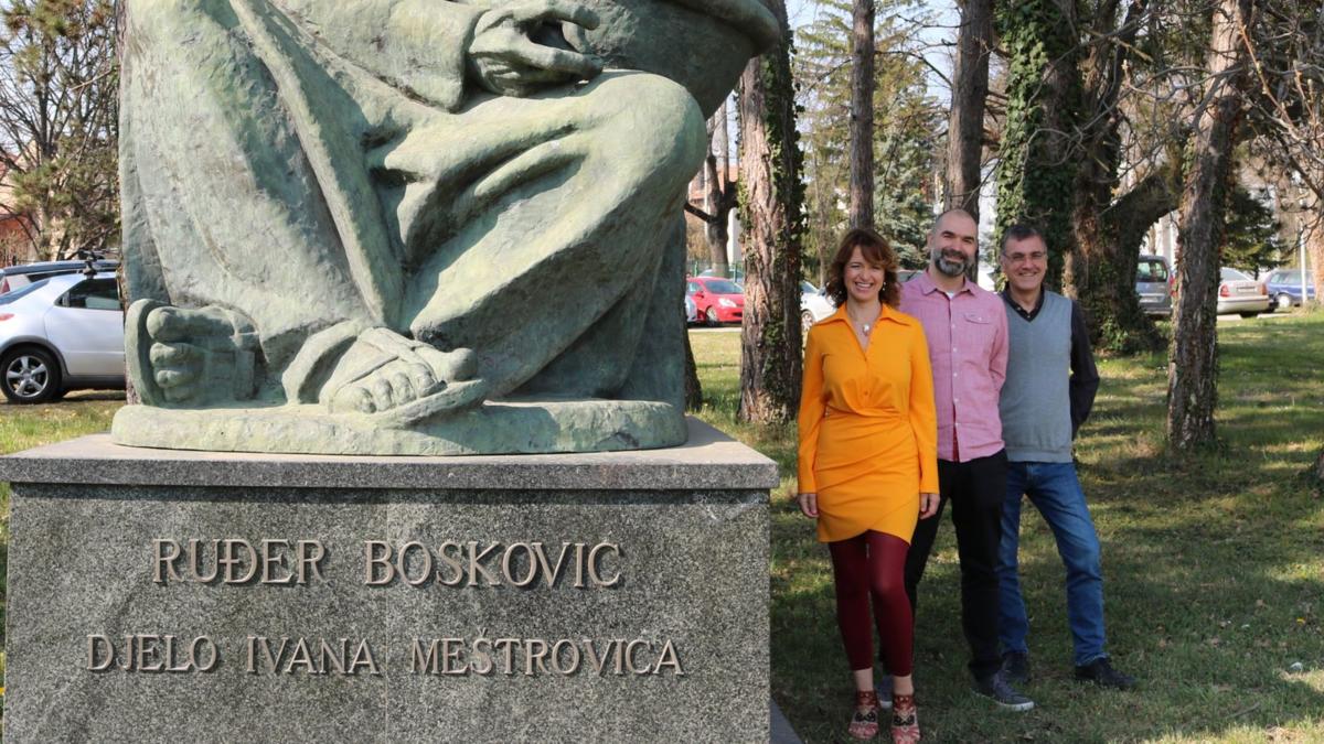
Iva Tolić, Marin Barišić and Nenad Pavin – Ruder Boskovic Institute
How are chromosomes organised during cell division?
Our bodies are made up of about one hundred trillion individual cells created by division from a single cell. Spindle fibers, dynamic micromachines composed of protein tubes – microtubules, are responsible for that division. Although the assembly of the spindle fiber is essential for proper chromosome division, this process has not been fully elucidated due to its complexity.
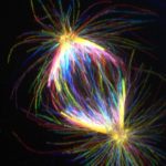
The spindle fiber has a unique architecture consisting of evenly distributed bundles of microtubules – kinetochore bundles, which are attached to chromosomes, and bridging bundles composed of microtubules, which connect the two poles of the spindle fiber by folding in the middle.
While the formation of kinetochore fibers has long been investigated in many laboratories around the world, how the bridging bundles are assembled has remained unknown. Without these bridging bundles, spindle fibers end in a star shape that cannot separate chromosomes. This is proof that bridging bundles play an indispensable role in cell division.
To clarify the formation of bridging bundles of microtubules, group of Professor Iva Tolić teamed up with the group of Professor Marin Barišić from the Danish Cancer Research Center in Copenhagen and the group of Professor Nenad Pavina from PMF Zagreb as part of the project of the Croatian Science Foundation (HrZZ) under the programme encouraging cooperation with Croatian scientists in the diaspora.
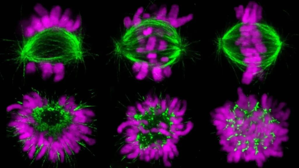
The project was completed with the publication of the results of interdisciplinary research in which, combining cell biology and theoretical physics, the researchers discovered the phase transition of microtubules from a sparse network structure to dense, well-separated, and properly organised bundles of the spindle fibers.
Experiments conducted by student Jurica Matković showed that this transition occurs because motor proteins on kinetochores, which are located in the central part of each chromosome, bind to microtubules.
Binding requires the activation of motor proteins by Aurora kinase B, a protein that has multiple roles during cell division. This result was proven by experiments based on motor protein mutants by researchers led by Prof. dr. sc. Marin Barišić.
When motor proteins bind to microtubules, cross-linking proteins cross-link the microtubules into a bundle attached to the kinetochore. The mutual repulsion of the condensed chromosomes, which push against each other on the equatorial plane of the spindle fibers and thus lead to the separation of the bundles to which they are attached and the expansion of the structure into a characteristic spindle shape, is responsible for not all microtubules joining into a single bundle. This novel mechanism of bundle formation is relevant not only to microtubule-based structures but also to cytoskeletal self-organisation in general.
Young researchers have developed new approaches and methods in cell microscopy
“To be able to reveal these complex processes, our research team devised new approaches and developed better methods for observing the spindle fibers. Rapid imaging of microtubules in the cross-section of the spindle fibers allowed us to monitor the dynamics of microtubule redistribution in a living cell. Until now, it was impossible to achieve this with conventional approaches due to the large number of microtubules and the high speed with which they are reorganised. With the help of super-resolution STED microscopy, young researchers were able to analyse the previously unseen architecture of microtubules in the earliest stages of cell division,” explains Professor Iva Tolić, head of research at IRB.
Super-resolution microscopy protocols were developed on a microscope purchased as part of a project financed from the European Regional Development Fund as part of the Operational Programme Competitiveness and Cohesion 2014-2020.
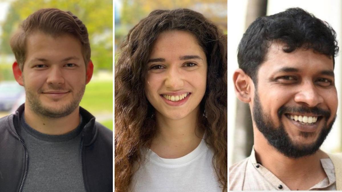
Jurica Matković, Mateja Ćosić and Subhadip Ghosh – Ruder Boskovic Institute
The group of Professor Nenad Pavina studied the physics of the formation of microtubule bundles from PMF, where postdoctoral student Subhadip Ghosh developed a theoretical model that allows for identifying the conditions necessary for the formation of the bundles. Since this is a complex process, a minimal model is helpful in understanding the interplay between microtubules, kinetochores, chromosomes, and crosslinker proteins and their roles in bundle formation. The model results support the central hypothesis that the attractive and repulsive mechanisms revealed in the experiments drive the formation of microtubule bundles.
The functional importance of this concept is evident in the context of proper chromosome division, given that PhD student Mateja Ćosić showed in this paper that improper formation of bundles leads to errors in chromosome division due to failure to correct improper connections between microtubules and kinetochore.
The authors propose the intriguing hypothesis that Aurora kinase B not only promotes the formation of overlapping bundles by activating motor proteins, but uses these same bundles as pathways to kinetochores to correct misconnections there.
Irregular and overly thin bundles lead to lagging of individual chromosomes, due to which one daughter cell may receive too many chromosomes, and the other too few.
“The wrong number of chromosomes is characteristic of tumor cells and is associated with the formation of metastases. That’s why errors during chromosome division are intensively investigated in laboratories around the world, and this work adds a piece to the puzzle of understanding cell division and diseases associated with this important process,” the scientists concluded.
For more, make sure to check out our dedicated Lifestyle section.